Fig. 1.
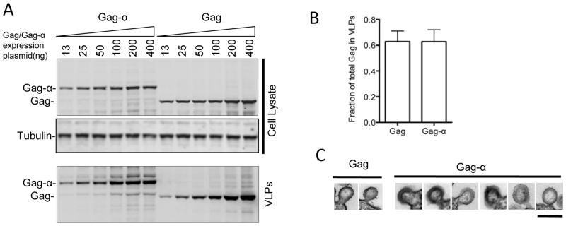
HIV-1 Gag-α is expressed and released as VLPs equivalently to Gag. (A) 293T cells were transiently transfected with increasing amounts of plasmids expressing either Gag or Gagα (13ng, 25ng, 50ng, 100ng, 200ng and 400ng). Cells were lysed 48 hours post-transfection and VLPs were prepared at the same time from culture supernatants by pelleting through a 20% sucrose cushion. Cell lysates and VLPs were subjected to SDS-PAGE and transferred onto nitrocellulose membranes. Western blots were probed with anti-Gag and anti-Tubulin antibodies. (B) The efficiency with which Gag and Gag-α are released from cells is shown as the fraction of the total Gag in 293T cell cultures that was present in extracellular VLPs following transfection with 500ng of plasmids expressing either Gag or Gag-α. Levels of Gag or Gag-α in VLPs and cell lysates was determined by quantitative western blotting. The mean and standard deviation of 7 experiments is plotted. (C) Gallery of thin-section electron micrographs showing viral like particles budding from 293T cells expressing Gag or Gag-α. Scale bar = 200nm.

