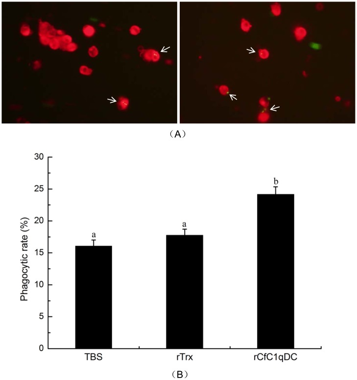
Figure 8. Phagocytosis enhanced by rCfC1qDC.
The hemocytes were resuspended with TBS buffer, rTrx in TBS buffer and rCfLec-1 in TBS buffer, respectively. Then E. coli was added into each hemocytes suspension for 45 min. The mixture was mounted onto a glass slide and incubation for 30 min to allow attachments of hemocytes to form a monolayer. After the fluorescence of nonphagocytosed bacteria was quenched by the trypan blue, hemocytes on each slide were counted (A). Phagocytic rate (PR) = (phagocytic hemocytes)/(total hemocytes)×100% (B). For each treatment, assay was performed in four different slides for statistic analysis. Bar = 10 µm.

