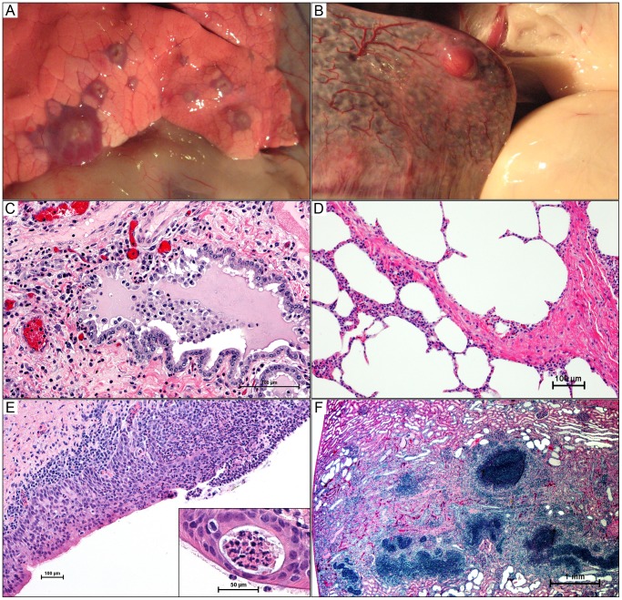
Figure 4. Gross and histologic lesions of caprine melioidosis.
A) Lung, multiple discrete subpleural targetoid pyogranulomas with tan purulent centers and consolidated hyperemic rims; B) a splenic subcapsular pyogranuloma bulging over the splenic surface with regional capsulitis and injected vessels; C) bronchiolar epithelium showing apical surface deciliation, segmental necrosis of the pseudostratified ciliated epithelium, neutrophil transcytosis, and luminal aggregates of neutrophils suspended in inflammatory edema; D) pulmonary septal thickening secondary to neutrophil infiltration; E) sever necrosuppurative/ulcerative tracheitis with neutrophil transcytosis and a mucosal abscess (inset); F) linear renal pyogranuloma extending from the cortex down into the medulla obliterating large areas of renal parenchyma. 4C–4F hematoxylin and eosin staining.

