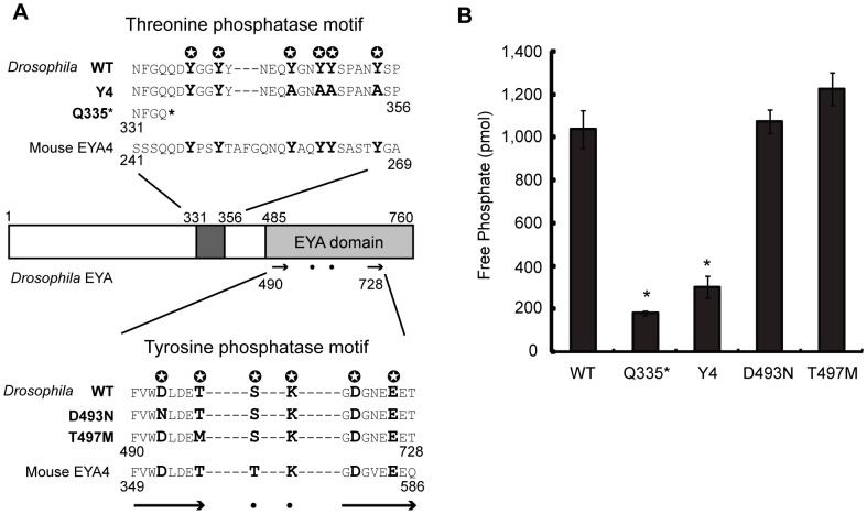
Figure 2. Threonine phosphatase domain in recombinant Drosophila EYA mutant proteins.
(A) Schematic view of Drosophila EYA with various mutant forms, WT, Y4, Q355*, D493N, and T497M. Two distinct phosphatase motifs are shown. Numbers indicate the positions of amino acid residues.  and bold letter indicate evolutionarily conserved amino acids in the motifs. Arrows and dots in the EYA domain correspond to those shown in the magnified view with amino acid sequences. Two motifs in amino acid sequences of Mouse EYA4 are shown. (B) Drosophila EYA threonine phosphatase activities of WT, Q355*, Y4, D493N, T497M were measured. Free phosphate in mol is indicated. One-way ANOVA was performed and followed by Dunnett's multiple comparison test. * indicates statistically significance (p<0.001) by comparing to WT.

