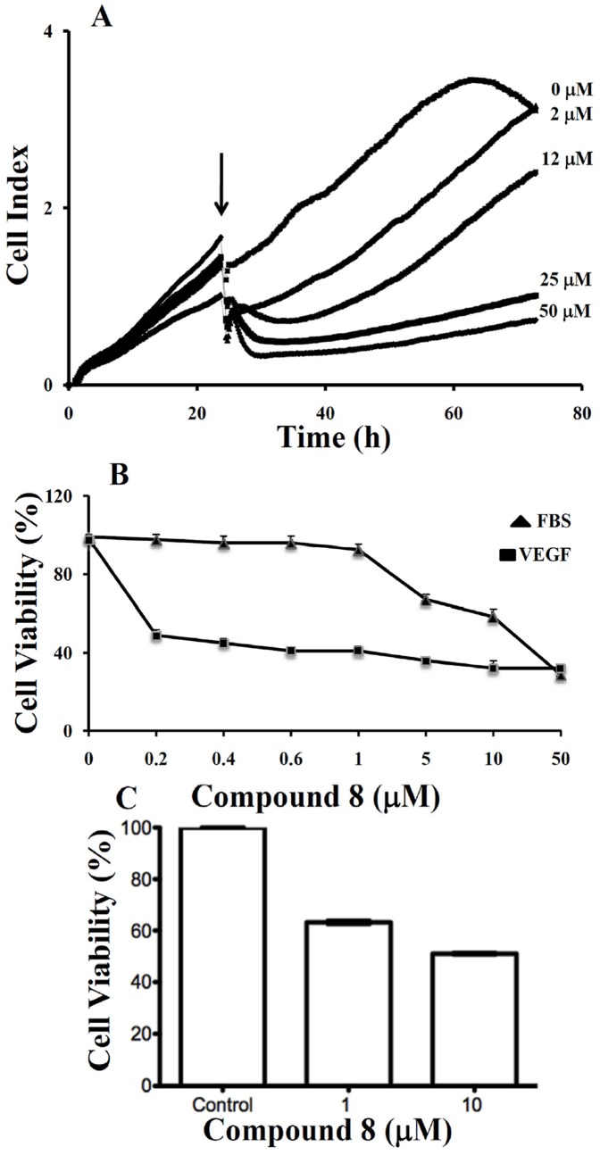
Figure 5. A. Real-time monitoring of the effects of compound 8 on the proliferation of LM8G7 cells.
Cells were seeded in ACEA's 96× e-plate™ at a density of 5×10³ cells per well, and continuously monitored using the RT-CES system up to 24 h, at which point Compound 8 (2 to 50 µM) was added. The cell index is plotted against time. The arrow indicates the time of the addition of compound 8. Data represent the mean values ± S.D. for three identical wells from three independent experiments. B, compound 8 poorly inhibited proliferation of UV♀2 under serum-replete conditions (FBS), but potently inhibited VEGF-dependent proliferation of UV♀2 cells as analysed by TetraColor One assay. Results were normalised to DMSO controls. Data represent mean values ± SD for three independent experiments. * P<0.05 versus control. ** P<0.01 versus control. C, compound 8 inhibits VEGF-stimulated proliferation of human vascular endothelial cells. HUVEC cells were seeded on 6-well plates at a density of approximately 1×10⁵ cells/well in M200 medium supplement with LSGS (Low serum growth Supplement). The next day cells were stimulated with 10 ng/mL of VEGF in the presence or absence of 1 µM and 10 µM compound 8. After 48 hrs, Alamar Blue was added directly into culture media at a final concentration of 10% and the plate was returned to the incubator. Optical density (OD) of the plate was measured at 540 and 630 nm. As a negative control, AB was added to medium without cells.

