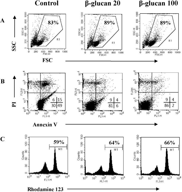
Fig. 3.
β-glucan protects splenocytes against spontaneous cell death. Splenocytes were cultured at a concentration of 5×10⁶ cells/5 ml in 6-well culture plates with 0, 20, or 100 µg/ml β-glucan treatment for 2 days. (A) The cell size (FSC/SSC) analysis, (B) annexin V-FITC/PI staining, and (C) Rhodamine 123 staining were performed by a flow cytometer. The number indicates cell percentage (A), the cell percentages in quadrants (B), and the cell percentage with high MFI (C), respectively. Each flow cytometric data set is presented from three independent experiments with similar results.

