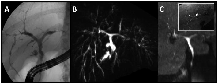
Figure 3.
29 year-old female with known PSC and ulcerative colitis. A multitude of strictures is depicted by T2w MRCP MIP (B) and ERCP (A). Gadoxetic acid-enhanced T1w MRC MIP (C) depicts the central ducts equally well. The MIP display in (C, inset: axial MIP) reveal the detail seen in the stack of axial images which revealed multiple stenotic and dilated segments to the 3rd (reader 1) or 4th (reader 2) branch.

