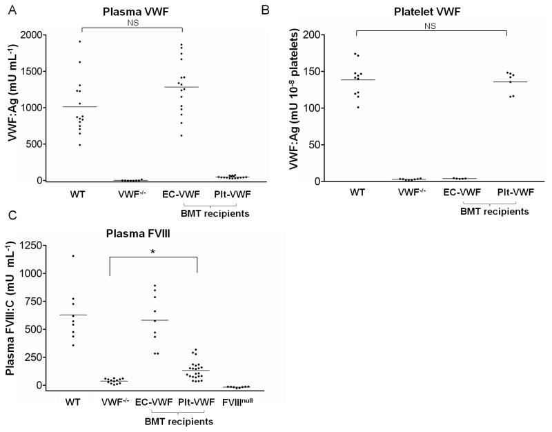
Fig. 1.
Murine models of EC-VWF and Plt-VWF established by crossed BMT. (A) Plasma samples and (B) platelet lysates collected from WT, VWF−/−, EC-VWF, and Plt-VWF mice were analyzed by ELISAto measure VWF antigen levels. EC-VWF mice had WT levels of plasma VWF, but no platelet VWF. Plt-VWF mice had WT levels of platelet VWF and a trace amount of plasma VWF. (C) FVIII activity in mouse plasma was quantified by chromogenic assay. FVIII:C of EC-VWF mice were similar to WT mice. Plt-VWF mouse plasma presented low FVIII:C but the levels were significantly higher than VWF−/− mice. NS, not statistically significant, *P < 0.05

