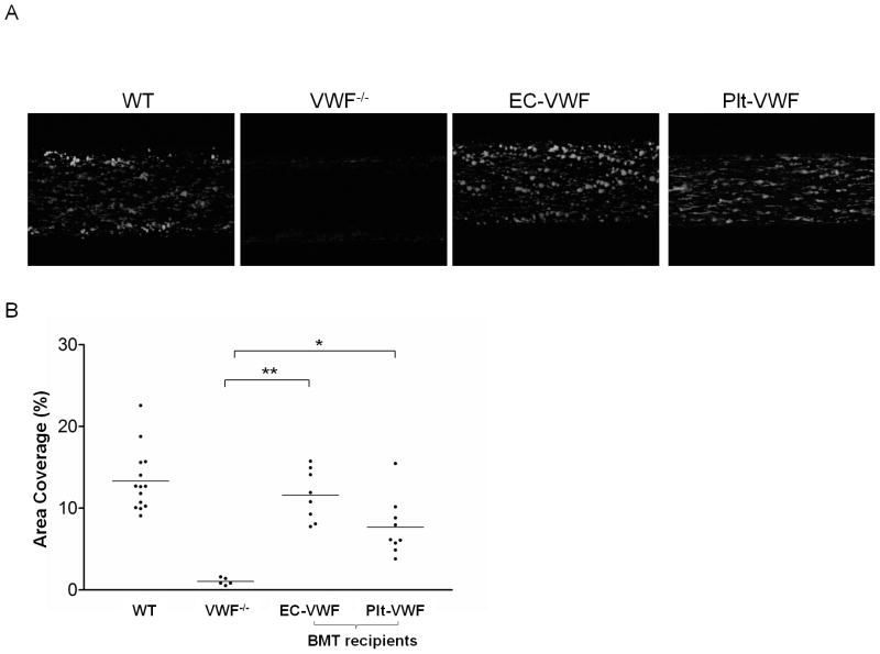
Fig. 2.
Platelet adhesion on type I collagen under flow. (A) Platelets in mouse whole blood from WT, VWF−/−, EC-VWF, and Plt-VWF were labeled with mepacrine and perfused over Vena8Fluor+Biochips coated with type I collagen at a shear rate of 2,000 s−1. Images were taken after 120 seconds of perfusion. (B) Percent of surface area covered by platelets after 120 seconds of perfusion was calculated using Metamorph software and plotted for each group of mice. Platelet adhesion of EC-VWF mouse blood samples to type I collagen were similar to WT mice. Surface coverage of Plt-VWF mouse blood samples was significantly higher than VWF−/− mice. *P < 0.05, **P < 0.001

