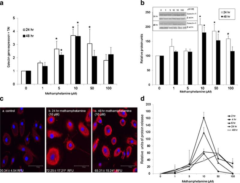
Fig. 1.
Methamphetamine regulates galectin-1 gene and protein expression in MDM. MDM were incubated with methamphetamine (1, 5, 10, 50, 100 μM), for 2, 4, 6, 24 or 48 h. a Gene expression for galectin-1 was determined using Q-PCR, (n=4). Data are expressed as transcript accumulation index (2–(ΔΔCT)) or TAI. b) Galectin-1 protein expression was determined using western blotting (n=4). Immunore-active protein bands were semi-quantified by densitometric analysis. Data are expressed as relative protein levels. Representative western blot for both galectin-1 and β-actin are shown in inset. c) Representative confocal image of galectin-1 protein expression (red) following methamphetamine (10 μM) incubation (a) control (b) 24 methamphetamine (c) 48 h methamphetamine with DAPI nuclear staining (blue). Relative fluorescent units (RFU) are shown in each panel (n=4, scale bar=75.49 μM). d) Supernatants were assayed for galectin-1 protein using ELISA (n=3). Data are expressed as relative protein units, with control value set at 0. Control cells release 120±5.2 pg/ml. All statistical significance was calculated using ANOVA followed by Bonferroni post-hoc test, * p<0.001; # p<0.05. • 2 h; ■ 4 h; ▲ 6 h; ◇ 24 h; X 48 h

