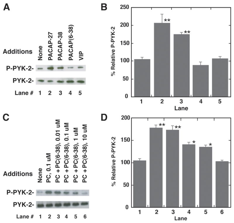
Fig. 3.

Phosphorylated PYK-2 and NCI-H838 cells. (A) PYK-2 tyrosine phosphorylation was determined 2 min after the addition of 100 nM PACAP-27, VIP, PACAP(6–38) or PACAP-38 (100 nM) to NCI-H838 cells (B) The mean value ± S.E. of 3 experiments is indicated; P < 0.01, ** using Student’s t-test. (C) The ability of varying doses of PACAP(6–38) to inhibit increases in PYK-2 tyrosine phosphorylation by PACAP-27 was investigated. (D) The mean value ± S.E. of 3 experiments is indicated; P < 0.05, *; P < 0.01, ** using Student’s t-test.
