Figure 4.

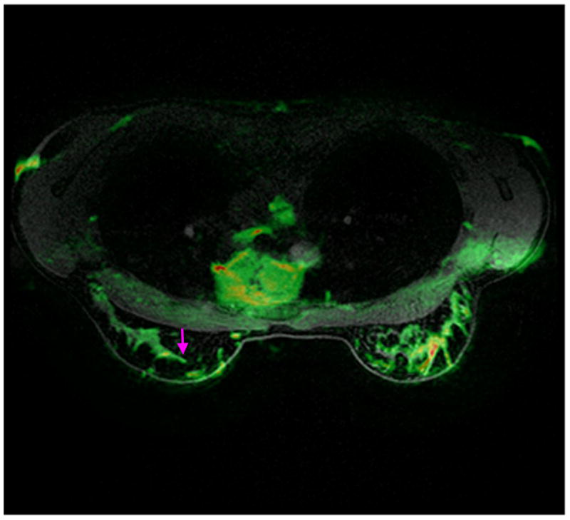
Mid-slice from a normal volunteer, with the anatomical and DWI images overlapped. The volunteer was shimmed using the best multi-plane shim strategy (strategy 7). Note the good match between the fine details of the two images (pink arrow).

Mid-slice from a normal volunteer, with the anatomical and DWI images overlapped. The volunteer was shimmed using the best multi-plane shim strategy (strategy 7). Note the good match between the fine details of the two images (pink arrow).