
Figure 2.
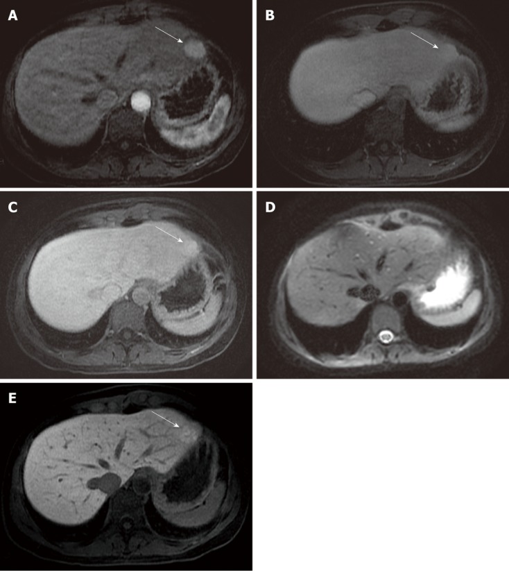
Focal nodular hyperplasia found in the left liver lobe. A: A round-shaped solid lesion (arrow) is depicted in the left liver lobe (in the II segment); B, C: The lesion (arrow) appears homogeneously hyperintense in the arterial phase, and remains slightly hyperintense in the portal and in the equilibrium phase; D: The lesion was missed on the diffusion image by both readers, probably due to its location near the gastric wall, along the liver surface; E: In the hepato-specific phase, the lesion (arrow) shows uptake of gadolinium-ethoxybenzyl-diethylenetriamine pentaacetic acid; a diagnosis of focal nodular hyperplasia was suggested due to the dynamic behavior observed after contrast administration.