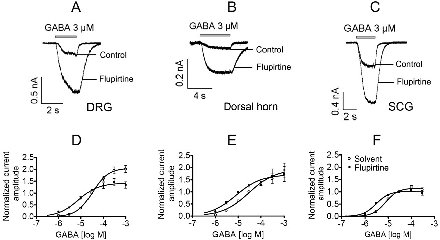
Figure 6.

Comparison of the effects of flupirtine on GABAA receptors in DRG, dorsal horn and SCG neurons. Neurons were clamped at −70 mV and increasing GABA concentrations were applied for 3 to 5 s in the presence of either solvent or 30 µM flupirtine. (A–C) Original traces evoked by 3 µM GABA in DRG, dorsal horn and SCG neurons, respectively. (D–F) Concentration–response curves obtained in DRG (n = 5 to 15), dorsal horn (n = 6 to 16) and SCG (n = 5 to 13) neurons. Amplitudes evoked by different GABA concentrations in solvent or flupirtine were normalized to those evoked by 30 µM GABA in solvent in the very same neuron. Values for statistical differences (F test) between EC50 values in the presence of either solvent or flupirtine are P < 0.001 for DRG, P < 0.05 for dorsal horn and P < 0.001 for SCG neurons, respectively.
