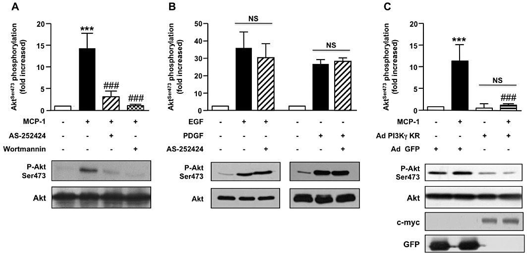
Figure 2.

PI3Kγ is required for PKB (Akt) phosphorylation in MCP-1-activated aortic SMC. (A, B) Pig aortic SMCs were pre-incubated (30 min) (A) with wortmannin (100 nM) or AS-252424 (100 nM) and stimulated with MCP-1 (10 ng·mL−1, 5 min) or (B) with AS-252424 and stimulated with EGF (10 ng·mL−1, 5 min) or PDGF (10 ng·mL−1, 5 min). Lysates were analysed by Western blot using antibodies against phosphorylated PKB (serine 473) or total PKB. Immunoblots from representative experiments and densitometry analyses are shown (means ± SEM, n= 4). (C) Pig aortic SMCs were infected with PI3Kγ KR-myc adenovirus (Ad PI3Kγ KR) or a GFP encoding adenovirus (Ad-GFP) as a control and stimulated with MCP-1 (10 ng·mL−1, 5 min). PKB phosphorylation and expression were analysed with antibodies against phosphorylated PKB (serine 473) or total PKB. Expression of PI3Kγ KR was analysed with anti-c-myc antibody and control adenovirus with anti-GFP antibody. Immunoblots from representative experiments are shown (n= 3). ###P < 0.001 versus MCP-1 alone and ***P < 0.001 compared with unstimulated cells.
