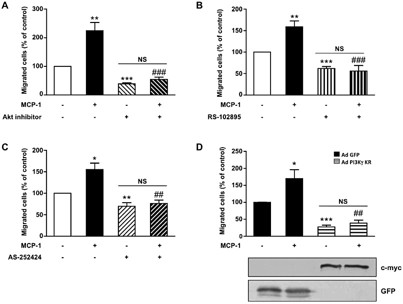
Figure 3.

PI3Kγ is essential for MCP-1-induced aortic SMC migration. Pig aortic SMC migration was quantified with a wound-healing assay using the Oris cell migration kit. Confluent SMCs were treated with MCP-1 (10 ng·mL−1) in serum-free medium and allowed to migrate in the wound surface for 48 h. Migrated cells were stained with DAPI and counted under a fluorescence microscope. Results are expressed as a percentage of control. (A) SMCs were treated with MCP-1 (10 ng·mL−1) only or together with PKB inhibitor (20 µM) in serum-free medium. (B) SMCs were treated with MCP-1 (10 ng·mL−1) alone or together with MCP-1 receptor inhibitor (RS-102895: 5 µg·mL−1). (C) SMCs were treated with MCP-1 (10 ng·mL−1) alone or together with a specific PI3Kγ inhibitor (AS-252424: 100 nM). (D) SMCs infected with GFP adenovirus (Ad-GFP) or with an adenovirus encoding a dominant negative form of PI3Kγ (Ad PI3Kγ KR) for 72 h were treated with MCP-1 (10 ng·mL−1). Expression of PI3Kγ KR was analysed with anti-c-myc antibody and control adenovirus with anti-GFP antibody. *P < 0.05, **P < 0.01, ***P < 0.001 compared with control, ##P < 0.01 compared with MCP-1 alone stimulated cells, ###P < 0.001 compared with MCP-1 alone stimulated cells.
