
Figure 4.
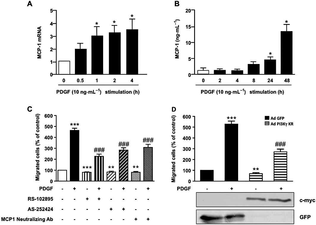
PI3Kγ amplifies PDGF-stimulated aortic SMC migration by an MCP-1/CCR2 pathway. (A) Pig aortic SMCs were stimulated with PDGF-BB (10 ng·mL−1) in serum-free medium for the indicated times. Total RNA was extracted and subjected to quantitative real-time PCR using specific primers for MCP-1. (B) Pig aortic SMCs were stimulated with PDGF-BB (10 ng·mL−1) in serum-free medium for the indicated times. Cell supernatants were analysed for MCP-1 protein content by a specific ELISA kit as described in Methods. (C) Confluent pig aortic SMCs were treated with PDGF-BB (10 ng·mL−1) alone or together with MCP-1 receptor inhibitor (RS-102895: 5 µg·mL−1) or a specific PI3Kγ inhibitor (AS-252424: 100 nM) or MCP-1-neutralizing antibody (5 µg·mL−1) in serum-free medium. Cells were allowed to migrate for 48 h and migrated cells were stained with DAPI and counted under a fluorescence microscope. Results are expressed as a percentage of the control. (D) Pig aortic SMCs infected with GFP adenovirus or with an adenovirus encoding a dominant negative form of PI3Kγ (PI3Kγ KR) for 72 h were treated with PDGF-BB (10 ng·mL−1) and analysed for migration as above. Expression of PI3Kγ KR was analysed with anti-c-myc antibody and control adenovirus with anti-GFP antibody. *P < 0.05, **P < 0.01, ***P < 0.001 compared with control, ###P < 0.001 compared with PDGF alone stimulated cells.