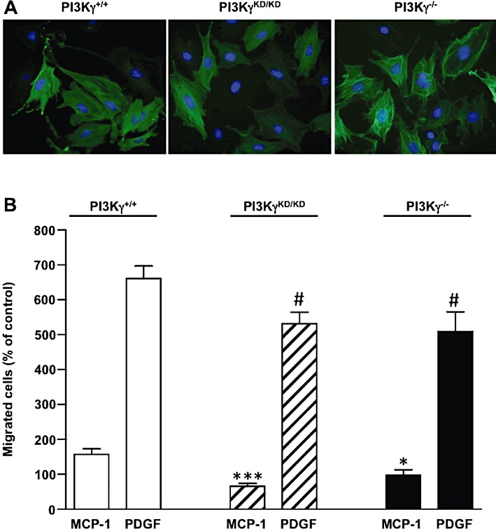
Figure 5.

Effect of PI3Kγ deficiency in MCP-1 and PDGF-induced migration of mouse aortic SMCs. (A) Aortic SMCs from wild-type (PI3Kγ+/+), PI3Kγ-deficient mice (PI3Kγ−/−) or mice expressing a catalytically inactive PI3Kγ (PI3KγKD/KD) were fixed and stained with an anti-smooth muscle α-actin antibody. Photomicrographs from representative experiments are shown. (B) Aortic SMCs from PI3Kγ+/+, PI3Kγ−/− or PI3KγKD/KD mice were stimulated with MCP-1 (10 ng·mL−1) or PDGF-BB (10 ng·mL−1) in serum-free medium and allowed to migrate for 48 h. Migrated cells were stained with DAPI and counted under a fluorescence microscope. Results are expressed as a percentage of PI3Kγ+/+, PI3Kγ−/− or PI3KγKD/KD non-stimulated cells, respectively. *P < 0.05, ***P < 0.001 compared with MCP-1-stimulated PI3Kγ+/+ cells; #P < 0.05 compared with PDGF-stimulated PI3Kγ+/+ cells.
