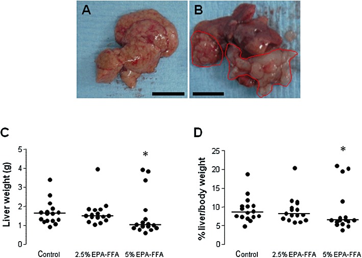
Figure 1.

Dietary EPA-FFA administration is associated with reduced growth of MC-26 mouse CRC cell liver metastases. (A,B) Macroscopic appearance of MC-26 mouse CRC cell liver metastases as either multiple small tumour foci throughout the liver (A; size bar = 10 mm) or a smaller number of larger, discrete tumours (highlighted in red) often involving the hilar region (B; size bar = 10 mm). (C) Individual liver weight values at killing. Bars represent the median value for each dietary group (control, n= 16; 2.5% EPA-FFA, n= 16; 5% EPA-FFA, n= 16). *P= 0.034 for the comparison between the 5% EPA-FFA-treated and control group; Kruskal–Wallis test. (D) Individual liver/body weight ratios at killing. Bars represent the median value for each dietary group (control, n= 16; 2.5% EPA-FFA, n= 16; 5% EPA-FFA, n= 16). *P= 0.13 for the comparison between the 5% EPA-FFA-treated and control group; Kruskal–Wallis U-test.
