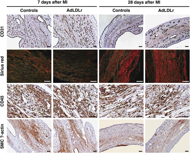
Figure 6.
(Immuno)histochemical analysis of the infarct area in control and AdLDLr-treated female C57BL/6 LDLr−/− mice 7 days and 28 days after MI. Representative photomicrographs show CD31-positive capillaries, Sirius red-stained collagen viewed under polarized light, CD45-positive leukocytes and smooth muscle cell α-actin-positive myofibroblasts. Scale bar represents 50 μm.

