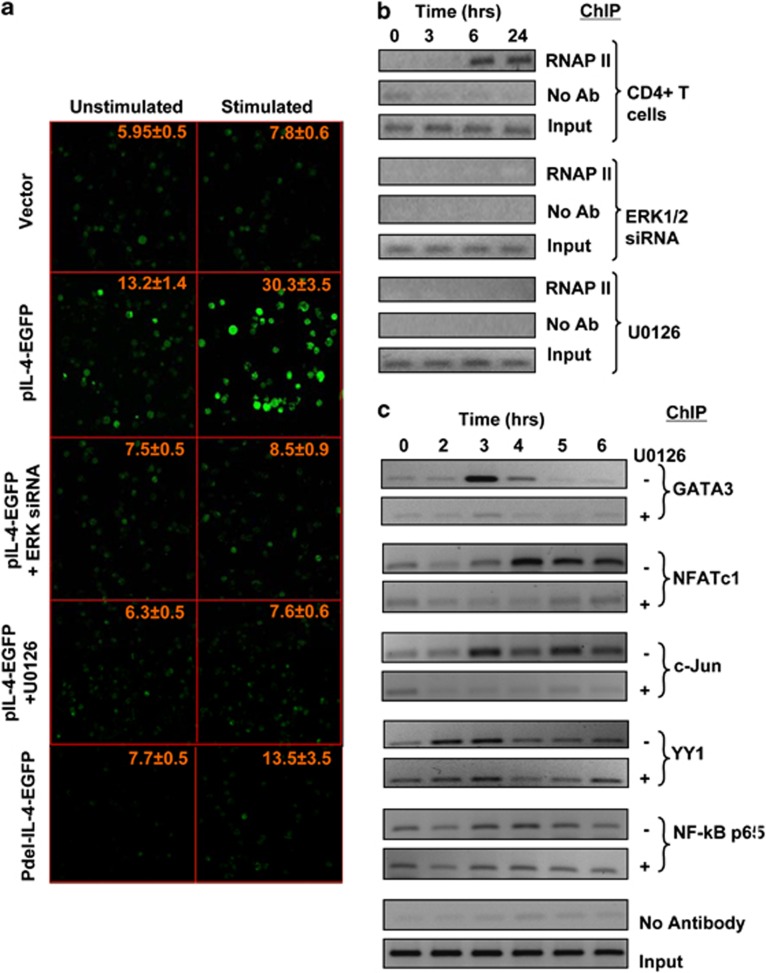
Figure 4.
Association of ERK is necessary for IL4 promoter activity. (a) Jurkat cells were transfected with the pEGFP1 vector (vector, top two panels), with the pIL-4-EGFP construct (from second to the fourth row of panels) or with a derivative of the pIL-4-EGFP construct in which the ERK-binding region was partially deleted (Pdel-IL-4-EGFP, bottom panels). This included two additional experimental groups in which pIL-4-EGF-transfected cells were also treated with either ERK-specific siRNA (panels in the third row from top) or with the MEK inhibitor U0126 (panels in the fourth row from top) as indicated. Figure shows the level of fluorescence obtained in each of these groups of cells when they were either left unstimulated or stimulated through the TCR for 24 h. Values indicated in each panel are the mean (±s.e.m) intensity of fluorescence measured for at least 40 cells from different fields in 3 separate slides for each group. (b) Naive CD4+ T cells were either left untreated (top panel) or treated with either ERK1/2 siRNA (KD) or U0126 (lower two panels). These cells were then stimulated through the TCR, and time-dependent recruitment of RNA polymerase II (RNAP II) to the IL4 promoter was monitored by ChIP analysis. The corresponding profiles obtained without anti-ERK antibody (no Ab) and input are also shown. (c) Time-dependent recruitment of indicated transcription factors, as determined by ChIP, to the IL-4 promoter after stimulation of cells through the TCR either in the presence (+) or absence (−) of U0126.

