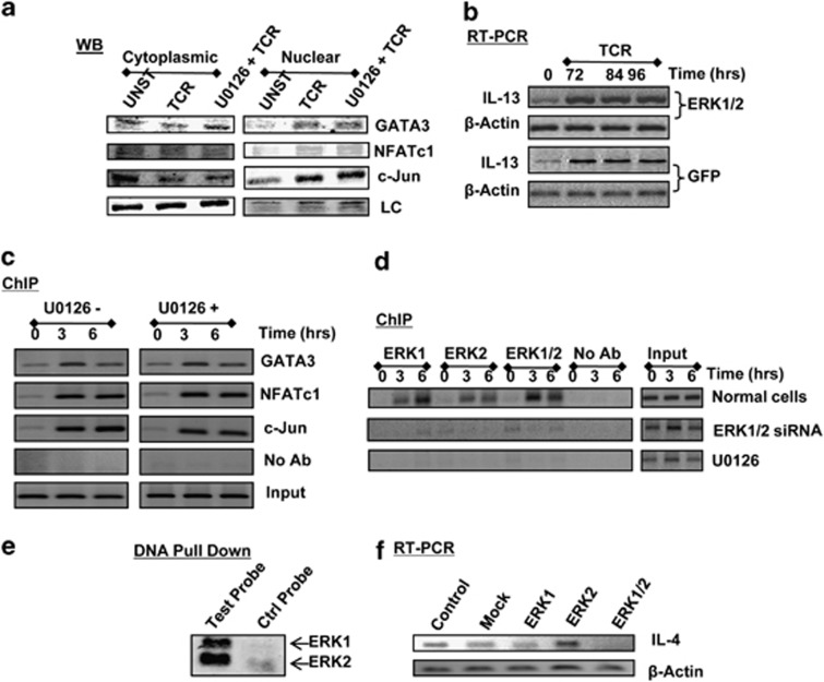
Figure 5.
The effect of ERK on IL4 gene expression is specific and involves both ERK-1 and ERK-2. (a) compares the distribution of three transcription factors (GATA3, NFATc1 and c-Jun) between the cytoplasmic and nuclear fractions in unstimulated (UNST), TCR-triggered (TCR, stimulation time of 30 min) and U0126-treated cells, followed by TCR triggering (U0126+TCR, stimulation time of 30 min). LC denotes the loading control, which was PLCγ2 for cytoplasmic fractions and Histone 1 for the nuclear fractions. (b) Effects of treatment of cells with either GFP or ERK1/2-specific siRNA on induction of the IL-13 transcript in activated cells. The time points at which transcript levels were determined by RT-PCR are indicated. Here, primers specific for β-actin were also included to provide an internal normalization control. (c) Results from a ChIP assay that monitored the recruitment of NFATc, c-jun and GATA3 to the region spanning −76 to −234 nt of the IL-13 promoter. Here, either untreated cells (U0126−) or cells treated with U0126 (U0126+) were stimulated through the TCR as indicated. Input and no Ab controls are also shown. (d) Results of experiments in which cells were activated through the TCR for indicated time and then subjected to a ChIP analysis using antibodies that were either specific only to ERK-1 or to ERK-2. For the purposes of comparison, an additional group was also included in which antibodies to both ERK-1 and ERK-2 were combined in the ChIP experiment (ERK1/2). Results shown are a representative of three separate experiments. (e) gives the results of a DNA pull-down experiment in which nuclear extracts from CD4+ T cells, stimulated through the TCR for 3 h, were incubated either with a 20-mer double-stranded DNA probe extracted from Figure 3d or with a control probe of similar length. In both cases, the probes were biotinylated. After incubation, the probes were separated by affinity chromatography (streptavidin-agarose), and the bound proteins eluted and analyzed for the presence of ERK-1 and ERK-2 by western blot analysis (see the ‘Methods' section). Results shown are from one of three separate experiments. (f) Results obtained in an RT-PCR experiment in which cells transfected separately with ERK1 siRNA (ERK1), ERK2 siRNA (ERK2) or with a combination of both (ERK1/2) were stimulated through the TCR. At the end of 84 h, cells were harvested, the total RNA was isolated and analyzed by RT-PCR for the IL-4 transcript. For the purposes of comparison, results obtained in cells that were either not treated with any siRNA (Control) or those treated with siRNA specific for GFP (Mock) are also shown. In all cases, primers specific for β-actin were also included to provide an internal normalization control.

