Figure 1.
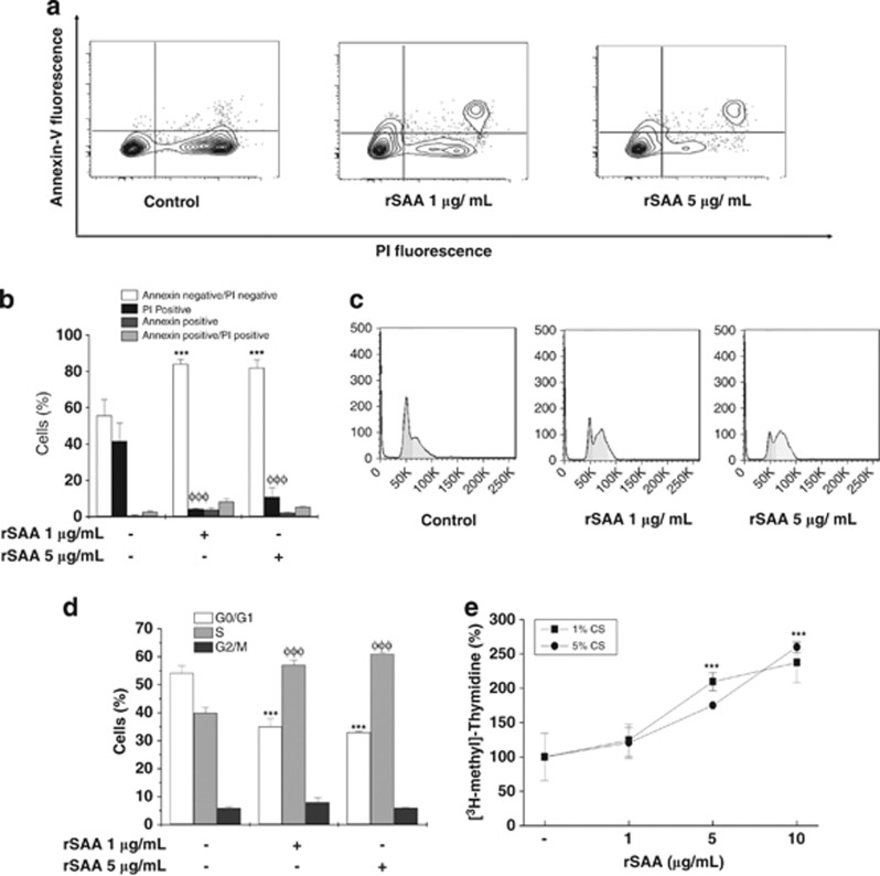
Enhanced cell viability and increased proliferation are induced by rSAA in 3T3-L1 preadipocytes. (a) Dot plots show the intensity of Annexin V fluorescence plotted on the Y-axis and PI fluorescence plotted on the X-axis. (b) The percentage of live cells (annexin-negative/PI-negative), necrotic cells (PI-positive), apoptotic cells (annexin-positive) and late apoptotic cells (annexin-positive/PI-positive) after analysis by flow cytometry. (c) Representative flow cytometry histograms showing cell cycle distributions. (d) The percentage of cells in each phase of the cell cycle stained with PI solution (2 μg ml−1). (e) 3T3-L1 cells were treated with increasing concentrations of rSAA (1–10 μg ml−1) in serum-deprived medium for 24 h. During treatment, the cells were labeled with [3H-methyl]-thymidine. Data are the mean±s.e. of three independent experiments, which were performed in triplicate and two-way analysis of variance were performed (***, φφφP<0.001 vs control groups).

