
Figure 1.
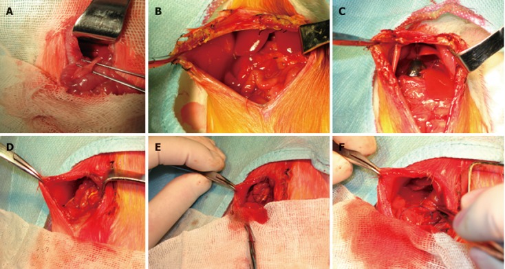
Images of experimental obstructive jaundice and internal biliary drainage. A: Dissection revealing the common bile duct; B: Reoperation after 3 d of common bile duct ligation. Light yellow abdominal ascites were present in the right side of the abdominal cavity; C: The proximal bile duct showed a remarkable expansion (dark blue color) after 3 d of common bile duct ligation; D: The PE-10 polyethylene tube was positioned with the end tied in the right hepatorenal recess; E: Brown bile flowed out while the catheter end was open; F: The distal 3 cm segment of the catheter was inserted into the duodenum for internal biliary drainage.