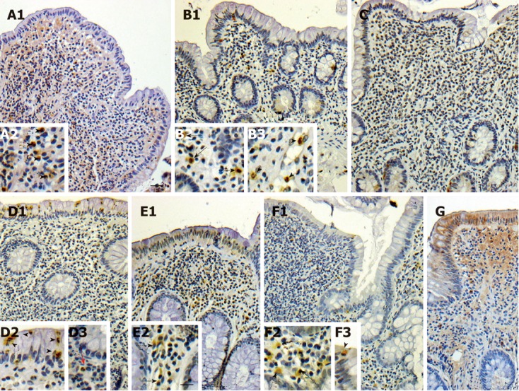
Figure 2.

matrix metalloproteinases-8 (A1), matrix metalloproteinases-9 (D1, E1), matrix metalloproteinases-12 (F1, G), matrix metalloproteinases-26 (B1) and tissue inhibitor of matrix metalloproteinase-1 (C) in pouch. Inset g’ added to figure from another sample, not shown in here in lesser magnification. Arrowheads depict neutrophils, black solid arrows plasma cells, black dotted arrows macrophages, red solid arrows eosinophils. Scale bars: 15 μm (A1, B1, C, D1, E1, F1,G); 7.5 μm (A2, B2, B3, D2, D3, E2, F2, F3). Stainings were performed using diaminobenzidine or NovaRED as chromogenic substrates and Mayer’s hematoxylin as counterstain. Images were obtained using a light-field microscope, and edited using Adobe Photoshop 7.0 (Adobe Systems Incorporated).
