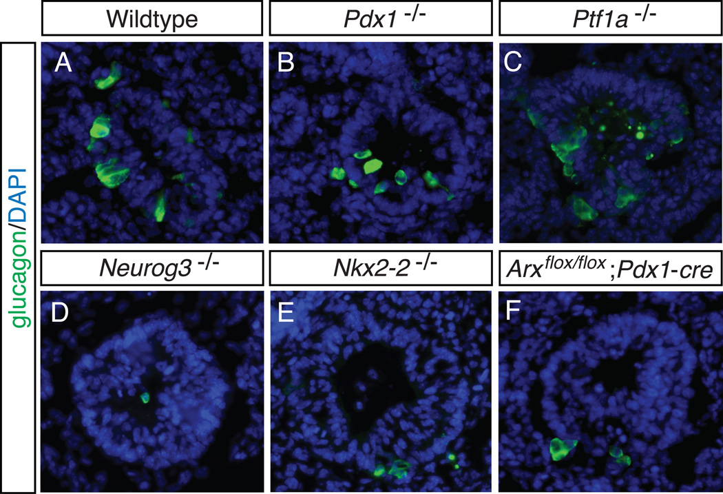
Figure 4. Early “first wave” glucagon-expressing alpha cells.
Early “first wave” alpha cells are present in mouse models despite the deletion of factors necessary for pancreas development and endocrine specification. Sagittal sections of E10.5 embryos from a representative wildtype littermate (A), Pdx1 null (B), Ptf1a null (C), Neurog3 null (D), Nkx2-2 null (E), and pancreas-specific deletion of Arx (F), were stained by immunofluorescence for the hormone glucagon (A–F). In all images glucagon-expressing cells are present. All images are of the dorsal pancreas. DAPI marks all nuclei. 40X

