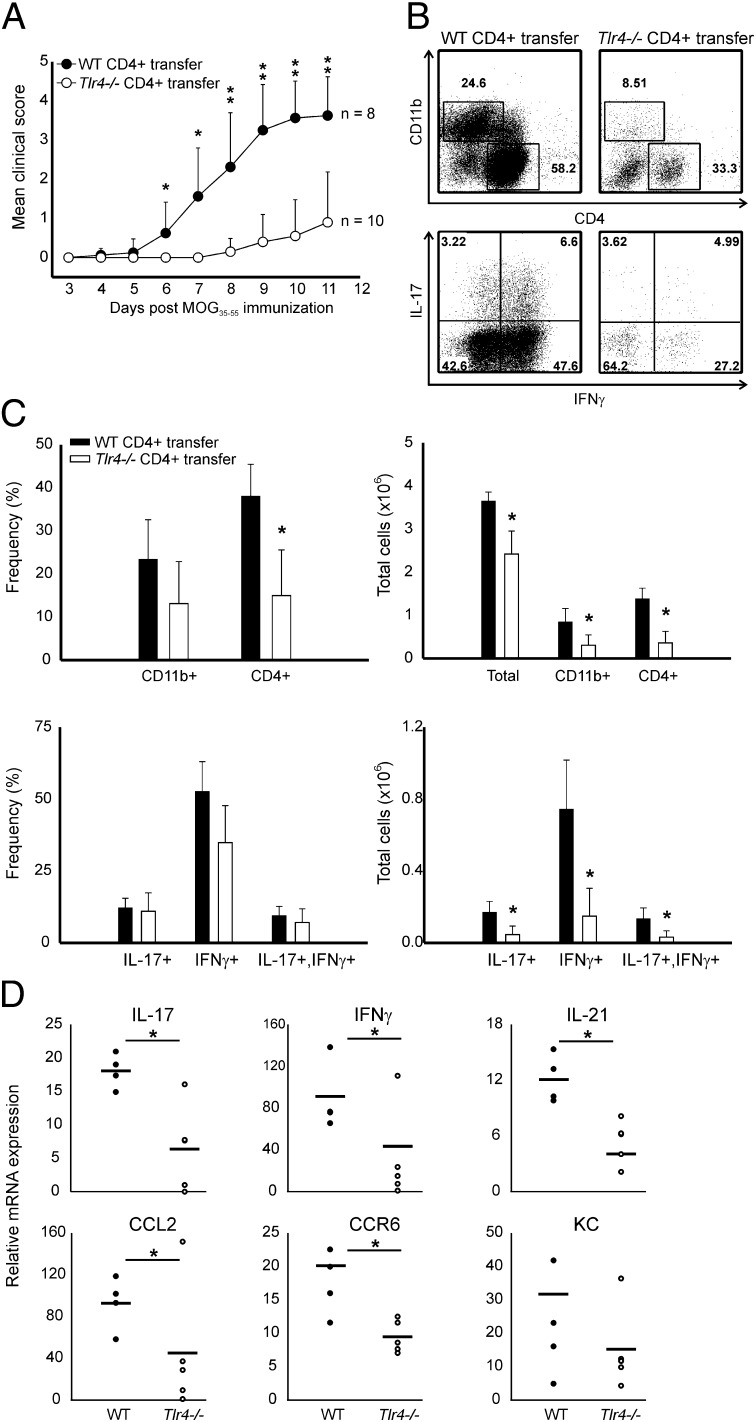
Fig. 3.
Tlr4−/− CD4+ T cells are defective in promoting EAE. (A) Representative clinical scores of Rag1−/− mice reconstituted with WT or Tlr4−/− CD4+ T cells. n = 8 for WT CD4+ T-cell transfer and n = 10 for TLR4-deficient CD4+ T-cell transfer. (B) Representative CNS staining of mice presented in A. CD4+ and CD11b+ cells were analyzed by surface staining (Upper). CD4+ T cells were analyzed for the production of IL-17 and IFN-γ following 6 h phorbol myristate acetate (PMA) restimulation (Lower). (C) Summary of CNS infiltration and cytokine production of the mice presented in A. (D) mRNA analysis of EAE brain and spinal cord tissues of Rag1−/− mice reconstituted with WT or Tlr4−/− CD4+ T cells. n = 5 mice per group. Data are representative of four independent experiments; *P < 0.05, **P < 0.001; Student's t test.

