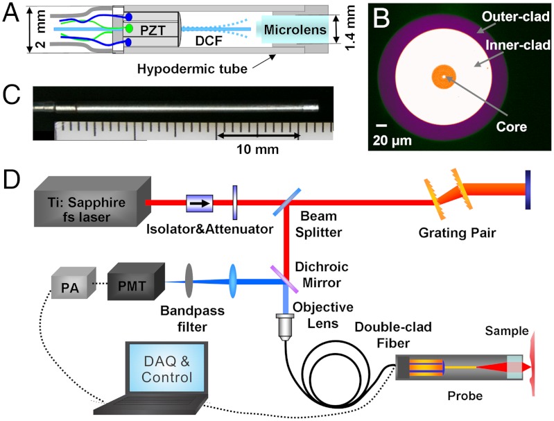
Fig. 1.
Optical design of the SHG endomicroscope and the imaging system schematic. (A) Schematic of the distal end of the scanning endomicroscope. PZT: tubular piezoelectric actuator; DCF: double clad fiber. (B) Photograph of the double-clad fiber cross-section. (C) Photograph of the assembled endomicroscope. (D) Layout of the endomicroscopy imaging system. PMT: photomultiplier tube; PA: preamplifier; DAQ: data acquisition unit.

