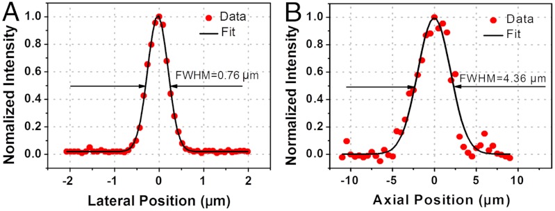
Fig. 2.
Endomicroscopy resolution measurements with two-photon excitation. (A, B) Fluorescence intensity profiles (dots) across the center of a 0.1-μm diameter fluorescent bead along lateral (A) and axial (B) direction. The fitted Gaussian curves are shown in black traces. The resolution was provided by the corresponding full-width-at-half-maximum (FWHM) of the Gaussian fit.

