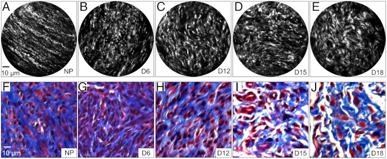
Fig. 4.
SHG endomicroscopy images of cervical tissues sections from nonpregnant (NP) (A) and pregnant mice at gestation days 6 (B), 12 (C), 15 (D), and 18 (E). Significant morphological changes in cervical collagen are evident over the course of pregnancy. Microscopic images of trichrome stained samples (F–J) at the same gestational time points show gross changes in collagen morphology.

