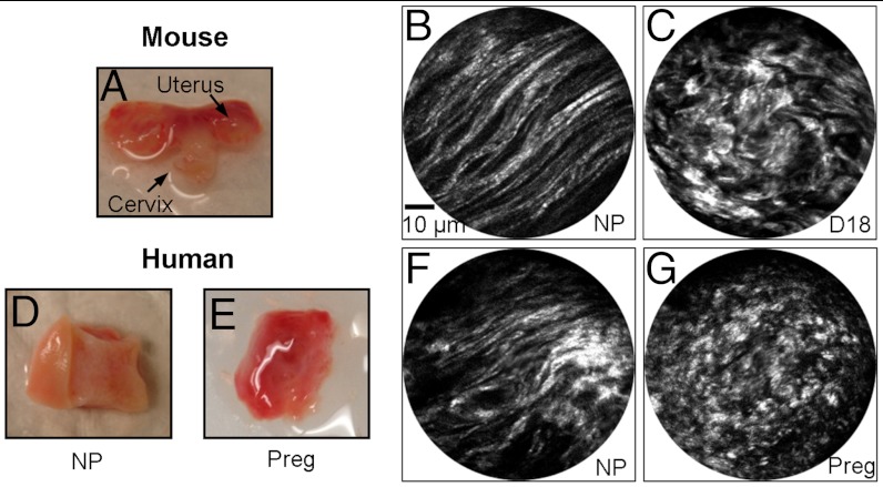
Fig. 6.
(A) Photo of an excised nonpregnant (NP) mouse cervix placed on a wax block. (B) and (C) are a representative SHG endomicroscopy image of NP and gestation day 18 mouse cervices, respectively, collected through intact epithelium. The SHG image quality and morphological features are similar to those observed from mouse cervix sections (see Fig. 4 A and E). (D) and (E) show photos of cervical specimens from NP and 36-wk pregnant women, respectively, with the exocervix facing up, and a representative SHG endomicroscopy image of these specimens are shown in (F) and (G), respectively.

