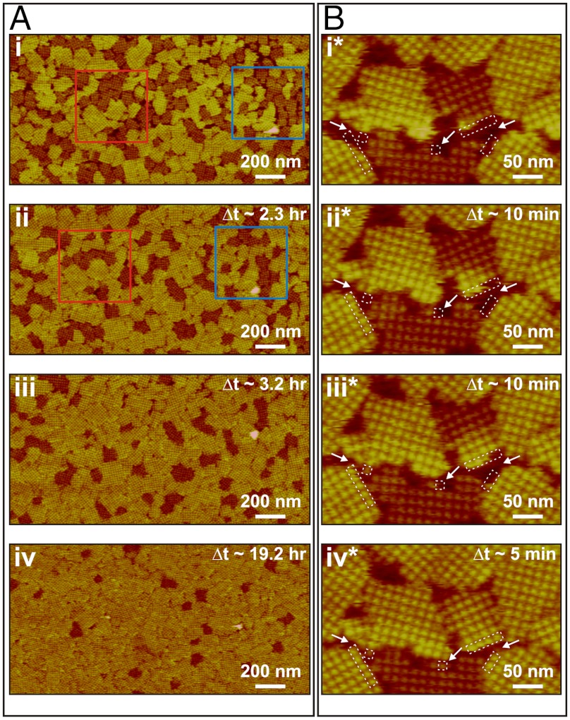
Fig. 3.
Sequences of in situ AFM images showing transformation from short to tall domains. Δt indicates time elapsed since collection of images i and i*. S-layer crystals were initially grown in 5 mL solution at 25 °C and further development of the domains was monitored in the same solution. Series in A shows evolution in distribution of domains in protein solution with CP = 41 μg/mL (10 mM Tris pH 7.2, 50 mM CaCl2, 100 mM NaCl) following an incubation time of 4.5 h. (i) Initial ratio of tall to short domains by area was 1.3. (ii) By Δt ∼ 2.3 h, about 25% of the short domains in i had transformed into tall domains without any dissolution of S-layer proteins from the domains. (iii) and (iv) At Δt ∼ 3.2 and 19.2 h, most of the short domains had transformed into tall domains. However, a few short domains still remained, indicating that the transformation does not go to 100% completion until much later times. Series in B shows transformation of a single domain in protein solution with CP = 70 μg/mL (10 mM Tris pH 7.2, 50 mM CaCl2, 100 mM NaCl) following an incubation time of 2.5 h. The transition began at the free edge of the short domain. In between (iii*) and (iv*), the short domain continued to grow by adding new tetramers at the bottom edge. Times of image collection were (i*) 2.5, (ii*) 2.7, (iii*) 2.8, and (iv*) 2.9 h.

