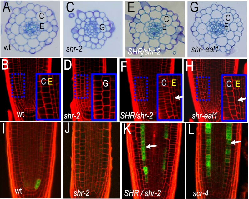
Fig. 1.
Roots with reduced SHR activity show ectopic periclinal cell divisions in the endodermis. (A, C, E, and G) Toluidine blue stained transverse cross-sections through 5-d-old roots; genotypes as indicated. (B, D, F, and H) Longitudinal confocal cross-sections through the root meristems of wild-type and mutant individuals; genotypes as indicated. Arrows point to precocious divisions. Region in the blue dashed box is magnified in the Inset. E, endodermis; C, cortex; and G, ground tissue. (I–L) Wild-type and mutant roots (as indicated) expressing pCYCD6;1:GFP-GUS. Arrows point to precocious periclinal cell divisions.

