Fig. 2.
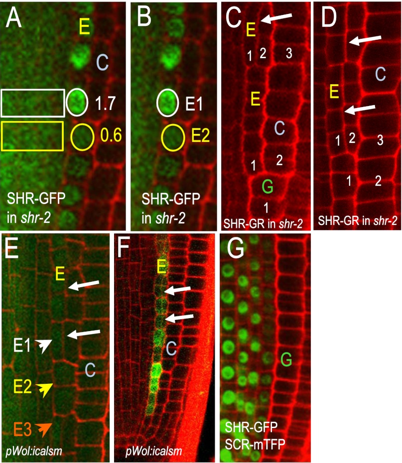
A reduction in SHR precedes cell division. (A and B) SHR-GFP in 14-d-old roots. Numbers in A indicate the endodermal-to-stele ratios of SHR-GFP fluorescence. White circles outline nuclei of recently divided endodermal cells, whereas yellow encircles those that are next in line to divide. (B) To eliminate the stele from the calculation of SHR-GFP levels in the endodermis, the fluorescence intensity in recently divided endodermal cells like E1 were compared with those that are in line to divide, E2. This comparison yielded an average ratio of 2.4. (C and D) Transfer of the shr-2 roots expressing pSHR:SHR-GR from dex-containing medium to standard MS medium lacking dex resulted in periclinal cell divisions in the endodermis (arrows). Small white numbers indicate the ground tissue layers relative to the stele. (E) Reduction in the movement of SHR-GFP in the pWOL:icalsm line resulted in periclinal cell divisions (arrows) and (F) activation of expression of pCYCD6;1:GFP-GUS in the endodermis. In E the white arrowhead E1 points to the nucleus of a divided endodermal cell, whereas the yellow E2 and orange E3 arrowheads point to undivided cells. (G) Expression of pSHR:SCR-mTFP in wild-type roots inhibits the movement of SHR-GFP and the formation of separate endodermis and cortex. E, endodermis; C, cortex; and G, ground tissue throughout.

