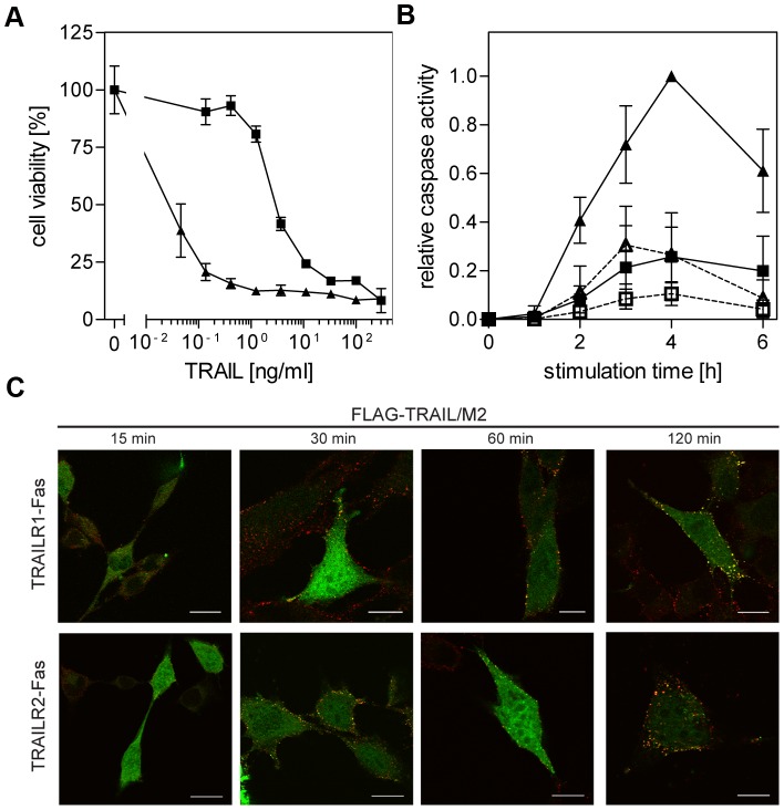
Figure 3. Differential TRAIL responsiveness, but comparable ligand/receptor cluster formation of TRAILR1-Fas and TRAILR2-Fas receptor chimeras.
A. Cytotoxic effects of TRAIL in TRAILR1-Fas and TRAILR2-Fas expressing mouse fibroblasts. MF-TRAILR1-Fas (closed squares) or MF-TRAILR2-Fas cells (closed triangles) were treated with serial dilutions of FLAG-TRAIL (previously cross-linked with anti-FLAG M2 antibody, 1 h 37°C) in the presence of cycloheximide (0.5 µg/ml). Cell viability was determined by crystal violet staining the next day. All experimental groups shown were performed in parallel, one representative experiment out of three is shown (performed in triplicates, shown are mean values ± SD). B. Caspase-8 (open symbols, dashed lines) and caspase-3 (closed symbols, solid lines) enzymatic activities following stimulation of MF-TRAILR1-Fas or MF-TRAILR2-Fas (squares and triangles, respectively) with 100 ng/ml FLAG-TRAIL (previously cross-linked with 2 µg/ml anti-FLAG M2 antibody, 1 h 37°C) were determined using specific fluorogenic substrates (Ac-IEPD-AMC and Ac-DMQD-AMC, respectively). Data points are mean values ± SD calculated from three independent experiments and normalized using the highest activity value (caspase-3 activity in MF-TRAILR2-Fas after 4 hours of stimulation). C. MF-TRAILR1-Fas and MF-TRAILR2-Fas cells were transiently transfected with a FADD-eGFP construct in the presence of 20 µM z-VAD-fmk. The following day cells were treated with FLAG-TRAIL (300 ng/ml, previously cross-linked with 2 µg/ml mouse anti-FLAG M2 antibody, 1 h 37°C) for the indicated time periods. After fixation cells were stained with an Alexa Fluor 546-labeled anti-mouse IgG antibody and examined by confocal laser-scanning microscopy. An optical section through the center of one representative cell for each time point is shown.

