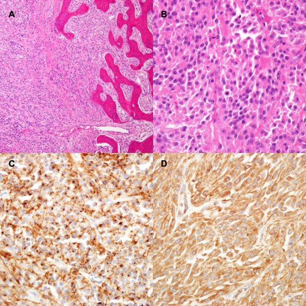
Figure 3.
Histopathology of the primary tumour. (A) Uniform cells with a sheet-like or vague parallel fascicle-like growth involving the nasal septum (×100, H&E). (B) Atypical cells having round-to-oval nuclei, with prominent nucleoli and occasional mitoses (×400, H&E). The tumour cells were diffusely positive for EMA (C, ×400) and vimentin (D, ×400).

