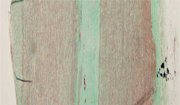
Figure 3:

Specimens were taken from the tailored double-layer convexity of the ascending aorta in a patient undergoing aortic valve replacement 6 years following isolated WA. Histology showed no sign of medial degeneration and perfect fusion of the two overlapped wall layers (Elastic van Gieson staining, 100× magnification).
