Figure 3.

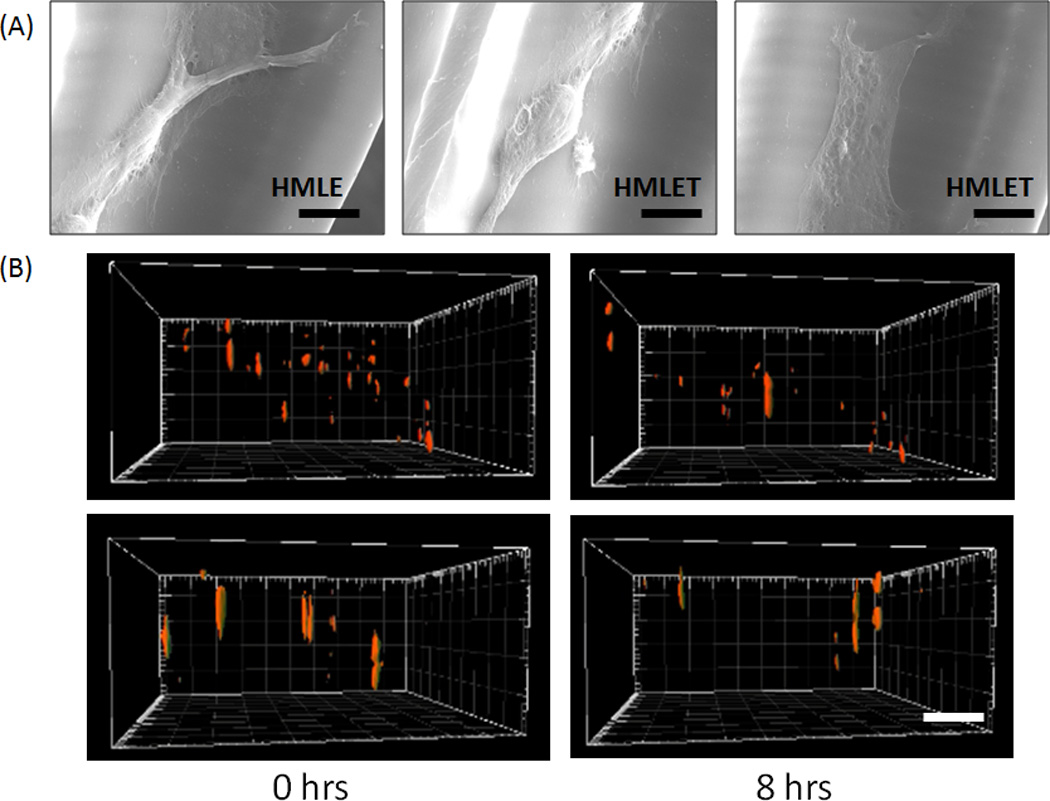
(A) Characteristic SEM images of HMLE and HMLET cells migrating inside the 3D log-pile scaffold. (Scale = 10µm). (B) Still images at 0 and 8 hours from reconstituted 3D confocal stacks of HMLE (top) and HMLET (bottom) on stiff 3D PEG log-pile scaffolds (Scale bar = 100µm).
