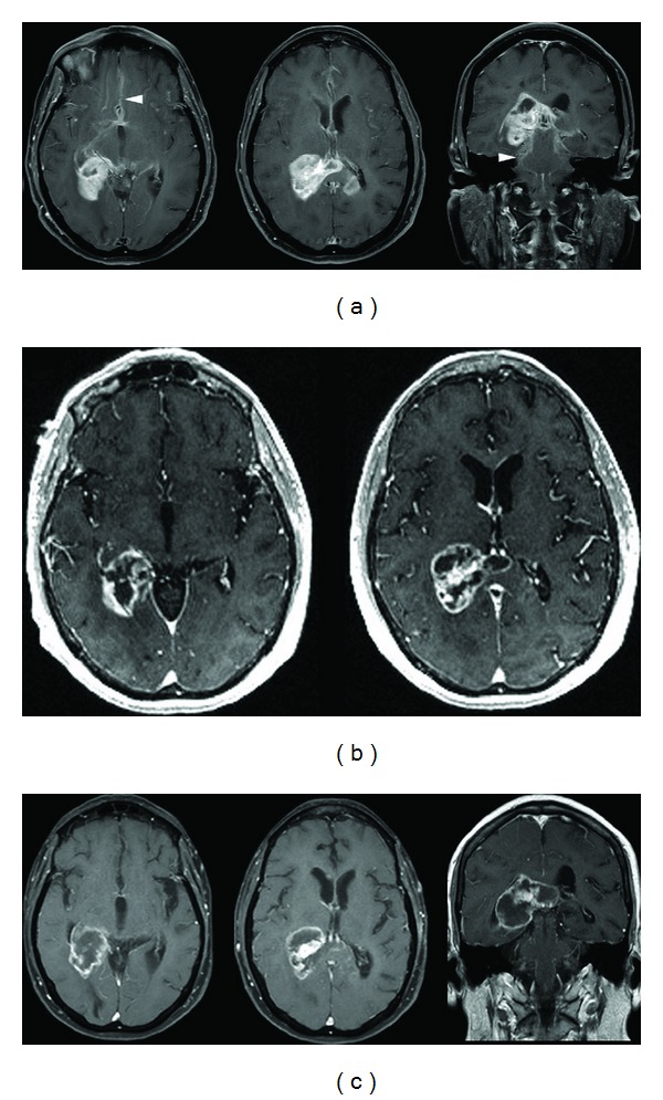
Figure 1.

(a) Initial contrast-enhanced axial and coronal T1-weighted fast spin echo (FSE) sequence demonstrating an avidly enhancing temporoparietal mass in a 57-year-old female patient who presented with short-term memory loss, headaches, subtle left-sided weakness, and unsteady gait. There is enlargement of the splenium with nodular enhancement within the contralateral corpus callosum. Extensive areas of subependymal and leptomeningeal enhancement (arrowheads) are present. (b) Contrast-enhanced axial spoiled gradient recalled (SPGR) sequence demonstrating overall decreased enhancement with formation of centrally necrotic areas after 5 days of corticosteroid therapy. The patient's improved functional status and the radiographic regression of the mass suggested a diagnosis of lymphoma. (c) Axial and coronal T1-weighted, contrast-enhanced FSE image obtained two weeks later showing increased nodular enhancement along the inferior and medial margins of the dominant mass and evolution of the necrotic areas. These changes suggested a diagnosis of glioma.
