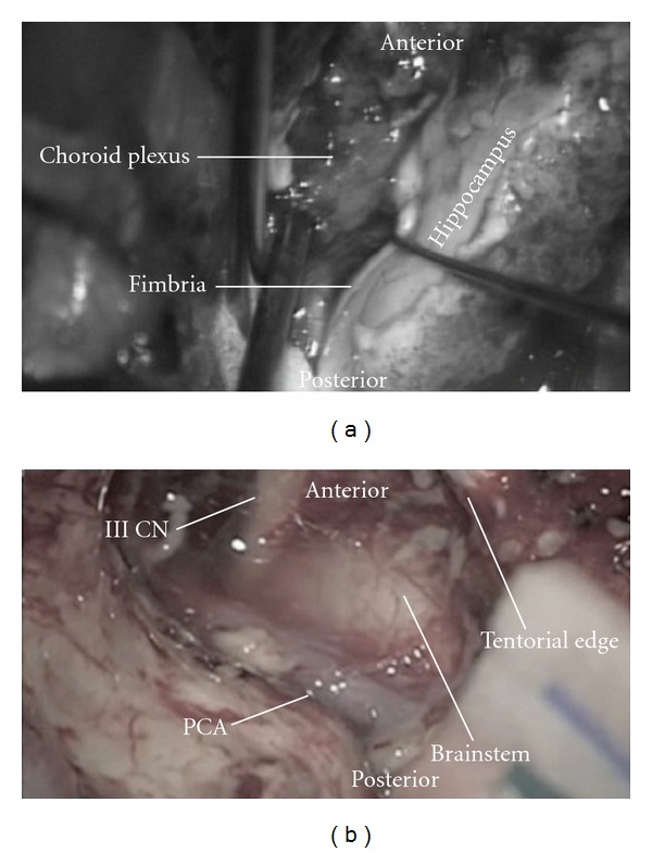
Figure 4.

Intraoperative photographs showing (a) the dissection of the fimbria to expose the choroidal point. (b) Postresection of the uncus and amygdala showing the third cranial nerve, brainstem, PCA (posterior cerebral artery), and the tentorial edge.
