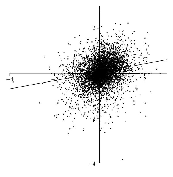
Figure 3.

A comparison of the intensity of protein spots, normalised to the intensity of the corresponding protein spot in control CNS, in Brodmann's area 46 from subjects with Sz (Y-axis) and bipolar disorder (X-axis). Spots in the upper left quadrant show increased levels in Sz compared to bipolar disorder whereas those in the lower right quadrant show increased levels of proteins in bipolar disorder with lower levels in Sz.
