Figure 2.

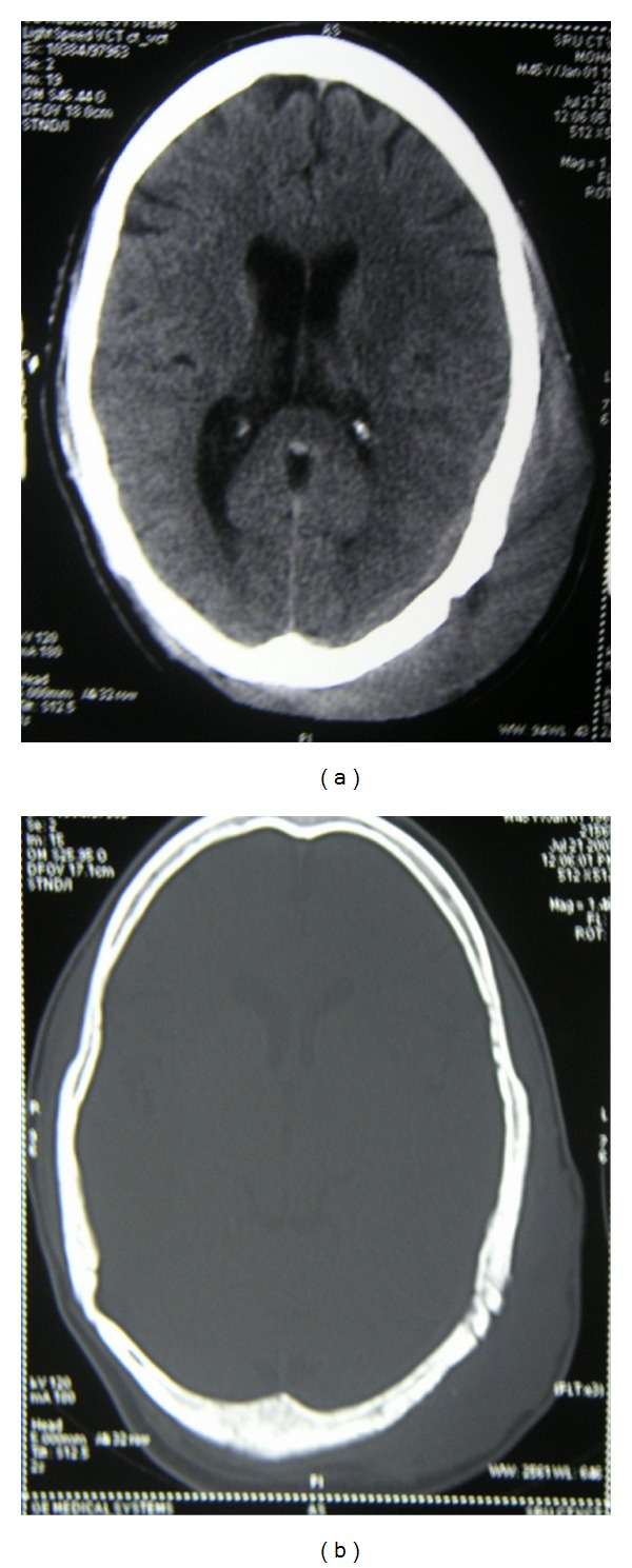
(a) CT scan axial section of the head showing scalp swelling in the left temporoparietal region after partial excision of the mass. (b) CT scan axial section showing scalp swelling after partial excision of the mass.

(a) CT scan axial section of the head showing scalp swelling in the left temporoparietal region after partial excision of the mass. (b) CT scan axial section showing scalp swelling after partial excision of the mass.