Figure 1.

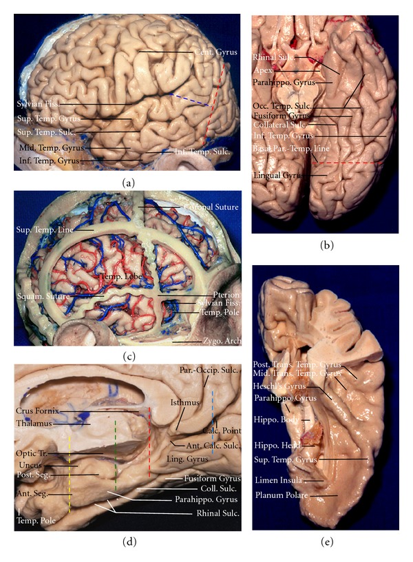
(a) Lateral view of the left hemisphere. The lateral surface of the temporal lobe consists of three parallel gyri: superior, middle, and inferior temporal gyri. These gyri are separated by the superior and inferior temporal sulci. The lateral parietotemporal line (red dashed line), an imaginary line connecting the preoccipital notch and parietooccipital sulcus, separates the temporal and occipital lobes, and the occipitotemporal line (blue dashed line), an imaginary line connecting the posterior margin of the sylvian fissure with lateral parietotemporal line, separates the temporal and parietal lobes. (b) Inferior view of the left temporal lobe. The basal surface of the temporal lobe consists of, from lateral to medial, the inferior margin of the inferior temporal gyrus, the fusiform gyrus, and the parahippocampal gyrus. The fusiform gyrus is separated laterally from the inferior temporal gyrus by the occipitotemporal sulcus, and medially from the parahippocampal gyrus by the collateral posteriorly and rhinal sulci anteriorly, which are not continuous in every case. The basal parietotemporal line connecting the preoccipital notch and inferior end of parietooccipital sulcus separates the temporal and occipital lobes at the basal surface. (c) The relationship of the temporal lobe with bony structures in a right hemisphere. The cranial sutures and the superior temporal line have been preserved, and the dura has been opened. The pterion is located at the lateral margin of the sphenoid ridge near the junction of the coronal, squamosal, and frontosphenoid sutures and the lateral end of the greater sphenoid wing and stem of the sylvian fissure. The squamosal suture follows the anterior part of the posterior limb of the sylvian fissure before turning downward, at the level of the postcentral and supramarginal gyri, to cross the junction of the middle and posterior third of the temporal lobe. The pole of the temporal pole fits into the cupped inner surface of the greater wing of the sphenoid bone. Most of the lateral surface of the temporal lobe is positioned deep to the squamous part of the temporal bone, except, the posterior part of the lateral surface extending deep to the parietal bone. The basal surface of the temporal lobe sits on the floor of the middle fossa and is positioned at the level of the upper edge of the zygomatic arch. (d) The medial view of the temporal lobe in a right hemisphere. This region is divided into three segments: anterior, middle, and posterior. The anterior segment begins at where the rhinal sulcus turns upward at the posterior edge of the temporal pole (yellow interrupted line) to a vertical line crossing the posterior edge of the uncus (green interrupted line), the middle segment extends from this point to the level of the quadrigeminal plate (red interrupted line), and the posterior segment extends from the quadrigeminal plate to the calcarine point (blue interrupted line) located at the junction of the parietooccipital and calcarine sulci. (e) The superior view of the left temporal lobe. This surface facing the the sylvian fissure is divided, from anterior to posterior, in three portion: the planum polare, the anterior transverse temporal gyrus, referred to as the Heschl's gyrus, and the planum temporale containing the middle and posterior transvers temporal gyri. Ant.: anterior; Cent., central; Calc.: calcarine; Fiss.: fissure; Hippo.: hippocampus; Inf.: inferior; Mid.: middle; Occ.: occipital; Parahippo.: parahippocampal; Par.-Occip.: parieto-occipital; Par.-Temp.: parietotemporal; Post.: posterior; Squam.: squamous; Sulc.: sulcus; Sup.: superior; Temp.: temporal; Trans.: transvers Tr.: tract; Zygo.: zygomatic.
