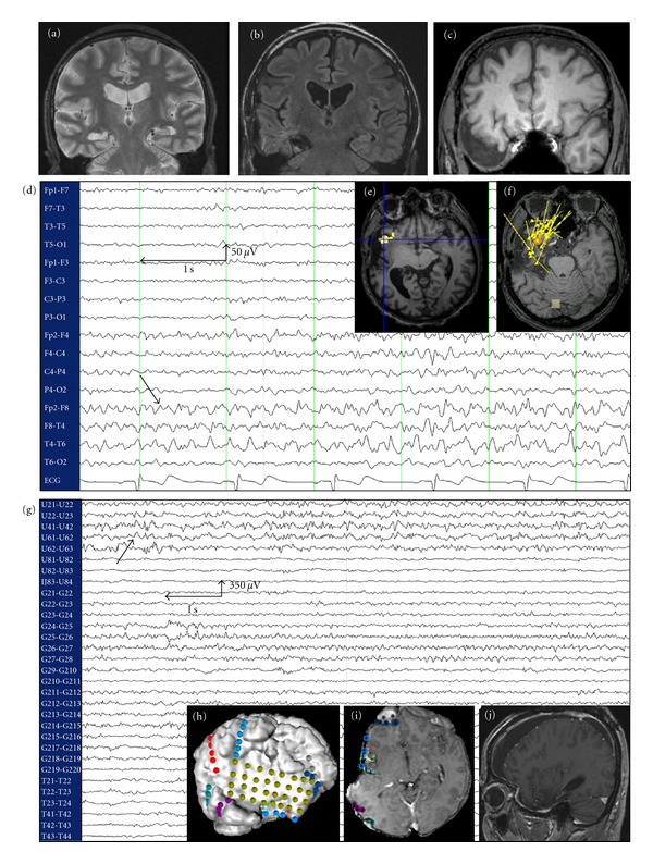
Figure 2.

Case of insular epilepsy masquerading as TLE operated at our institution. (a) MRI showing right hippocampal sclerosis in a 52-year-old patient with medically refractory epilepsy since age 20. (b) Presurgical evaluation led to a selective amygdalohippocampectomy that failed to control seizures. (c) Temporal lobectomy was subsequently performed, but the patient still continued to have seizures. (d): Postlobectomy scalp EEG continued to show ipsilateral temporal epileptiform activity. (e) and (f) fMRI-EEG and MEEG, respectively, localized the epileptogenic zone to the ipsilateral right insular cortex. (g) Intracranial EEG identified an epileptogenic focus in the anterior insular cortex. (h) and (i) Implantation of the intracranial electrodes. (j) MRI following partial anteroinferior insulectomy. The patient has been seizure-free since the operation (followup: 6 months).
