Figure 2.

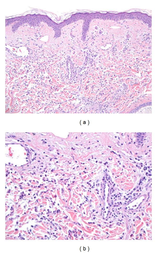
(a) Low magnification showing a diffuse dermal infiltrate of neutrophils accompanied by leukocytoclastic debris. (b) High magnification highlighting the neutrophils.

(a) Low magnification showing a diffuse dermal infiltrate of neutrophils accompanied by leukocytoclastic debris. (b) High magnification highlighting the neutrophils.