Figure 1.

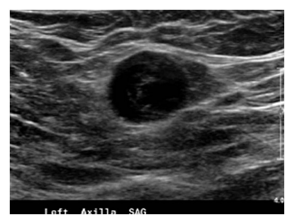
Ultrasound image at the time of core needle biopsy shows a well-circumscribed, 1.7 × 1.5 × 1.4 cm mass in the left axilla.

Ultrasound image at the time of core needle biopsy shows a well-circumscribed, 1.7 × 1.5 × 1.4 cm mass in the left axilla.