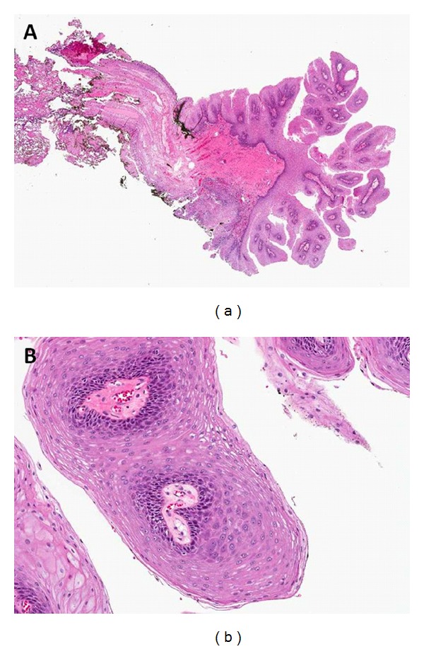
Figure 2.

Low power (10×) and high power (50×) hematoxylin and eosin stained sections from the uvula lesion. (a) The low power view demonstrates a papillary lesion consisting of multiple squamous lined papillary fronds containing fibrovascular cores. (b) At higher power the squamous cells show bland histological features. These findings are characteristic of a squamous papilloma.
