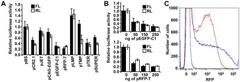
Figure 3. Effects of co-transfected plasmids on expression of luciferase reporters.
(A) Different plasmids have different effects on luciferase reporters. HEK-293 cells were co-transfected with 100 ng/well of each luciferase reporter and 150 ng of a tested plasmid. Renilla luciferase (RL) and firefly luciferase (FL) activities in pBS co-transfection were set to one. Data represent results of four transfection experiments performed in triplicates. Error bars = SEM. (B) Dose-dependent suppression of luciferase activities by co-transfected pEGFP-C1 (upper panel) and pRFP-T (lower panel). HEK-293 cells were co-transfected with 100 ng/well of each luciferase reporter and 0–250 ng/well of pEGFP-C1 or pRFP-T. The amount of transfected DNA was kept constant by adding pBS. Error bars = SEM. Data represent results of four transfection experiments performed in triplicates. (C) pEGFP-C1 negatively affects RFP reporter expression. HEK-293 cells were co-transfected with 150 ng/well of pCI-RFPT plasmid and 350 ng/well of pBS or pEGFP-C1 plasmid. RFP expression was analyzed 36 hours post-transfection by flow cytometry. X axis = RFP fluorescence intensity. Y axis = cell count. Colored curves show distribution of RFP signal as follows: black curve = untransfected cells; blue curve = pCI-RFPT + pBS co-transfection, and red curve = pCI-RFPT + pEGFP-C1 co-transfection. Total counts of transfected (RFP-positive) cells were identical in both samples (Fig. S1C). The shape of the red curve suggests that pEGFP-C1 reduces RFP fluorescence in transfected cells. The experiment has been performed three times, results from a representative experiment are shown.

