
Figure 2. NOL11 localization is dependent on RNA polymerase I transcription.
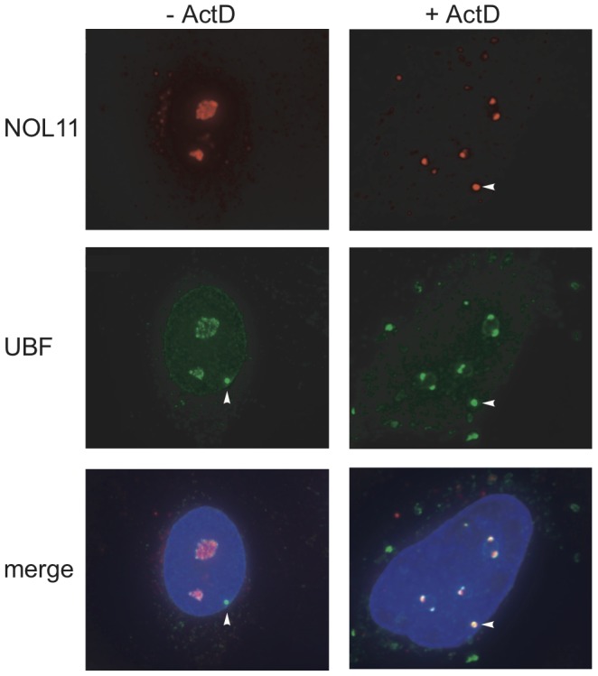
NOL11 was detected in HT1080-3D1 cells using an anti-NOL11 antibody. An anti-UBF antibody was used to stain nucleoli and pseudo-NORs. Co-localization of NOL11 and UBF is shown in the merged image. DAPI was used to stain chromatin. Cells were either not treated (−ActD) or treated (+ActD) with 0.1 µg/ml ActD for 1 h before fixation. Blue = DAPI, Red = NOL11, Green = UBF. Pseudo-NORs are indicated with arrowheads.