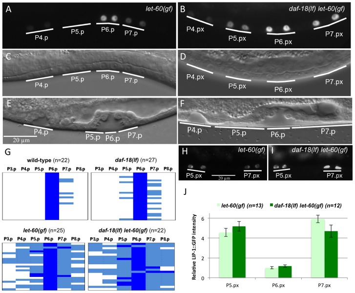
Figure 1. DAF-18 inhibits 1° vulval fate specification.
(A) Expression of the 1° cell fate marker EGL-17::CFP in let-60(gf) single and (B) daf-18(lf) let-60(gf) double mutants at the Pn.px stage. (C and D) show the corresponding Nomarski images of the animals shown in (A) and (B), respectively. (E) Morphology of the vulval invagination in a let-60(gf) mutant at the Pn.pxxx stage. Note that the descendants of P5.p and P7.p remain attached to the cuticle, which is characteristic of the 2° cell fate. (F) P5.p and P7.p often detach from the cuticle in daf-18(lf) let-60(gf) animals, resulting in a broad invagination with abnormal morphology. (G) Quantification of the 1° fate marker EGL-17::CFP expression at the Pn.px and Pn.pxx stages in the indicated genetic backgrounds. Dark blue represents cells with high EGL-17::CFP expression, corresponding to at least 50% of the intensity observed in the P6.px(x) cells, light blue represents cells with clearly visible but less than 50% of the P6.px(x) signal, and white spaces represent cells with undetectable levels of EGL-17::CFP. (H) Expression of the 2° fate marker LIP-1::GFP in let-60(gf) and (I) daf-18(lf) let-60(gf) animals. (J) Quantification of LIP-1::GFP expression in the P5.p through P7.p descendants. Signal intensities are indicated relative to P6.px(x) in let-60(gf).

